Types, incidental detection, partial nephrectomy, ablation, targeted therapy, and immunotherapy
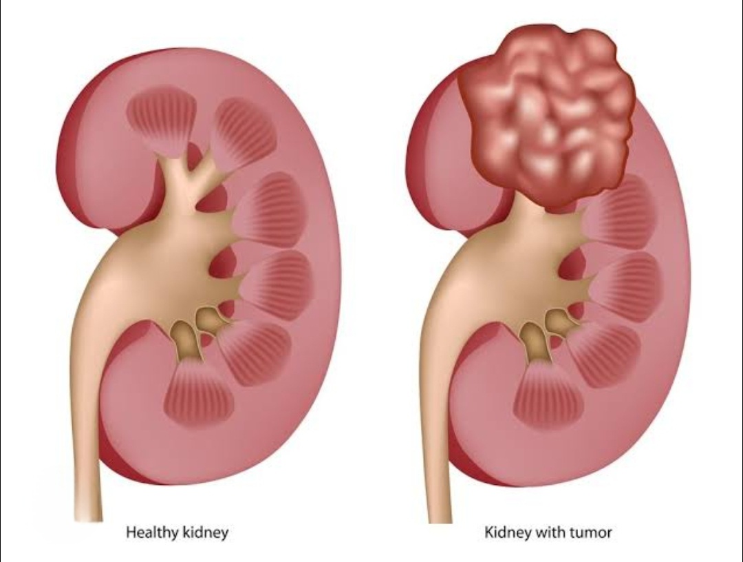
Renal cell carcinoma (RCC) is the most common kidney cancer, arising from tubular cells. Clear cell RCC accounts for 70-80%. Other types: papillary (15%), chromophobe (5%). The classic triad (flank pain, hematuria, mass) now occurs in <10% of cases — most are detected incidentally on imaging.
About 70% of kidney cancers are found incidentally on ultrasound or CT done for unrelated reasons. When symptomatic, patients may notice blood in urine, flank pain, or a palpable mass. Weight loss, fatigue, and fever may occur in advanced cases. Paraneoplastic syndromes (hypertension, polycythemia, hypercalcemia) occur in 20%.
CT with contrast (triphasic protocol) is the primary imaging tool — it characterizes the mass, determines extent, and assesses lymph nodes and veins. MRI adds detail for vein invasion. Chest CT detects lung metastases. Biopsy is not always needed pre-operatively but is used for small masses when ablation is planned or metastatic disease is suspected.
Partial nephrectomy (nephron-sparing surgery) removes only the tumor, preserving the remaining kidney. It is the standard of care for T1 tumors (<7 cm). Robotic-assisted partial nephrectomy offers precise tumor excision with minimal blood loss. Preserving kidney function reduces long-term cardiovascular risk and is especially important in patients with a single kidney or CKD.
Radiofrequency ablation (RFA) and cryoablation use heat or cold to destroy small tumors (<3 cm) percutaneously. Performed under CT or ultrasound guidance under local anesthesia. Oncologic outcomes are comparable to surgery for small tumors in elderly or surgically unfit patients. Local recurrence rate is 5-10%, higher than surgery.
Have more questions? Book a consultation with Dr. Samer Morsy
Radical nephrectomy removes the entire kidney and is used for large tumors (>7 cm), tumors in challenging locations, or vein thrombus cases. Laparoscopic and robotic approaches minimize recovery time. The adrenal gland is removed only when involved. Lymph node dissection is performed for staging when nodes appear enlarged on imaging.
Clear cell RCC is treated with VEGF-targeted therapy (sunitinib, pazopanib, cabozantinib) and immune checkpoint inhibitors (nivolumab + ipilimumab or pembrolizumab + axitinib). Combination immunotherapy is now first-line. Cytoreductive nephrectomy (removing the primary tumor) benefits selected patients with metastatic disease.
Small renal masses (<2 cm) in elderly patients can be monitored with active surveillance (serial imaging every 6-12 months) rather than immediate intervention. Growth rate under 3 mm/year is reassuring. The risk of metastasis for masses under 2 cm is under 2%. This avoids over-treatment in patients with limited life expectancy or high surgical risk.
Stage I (T1): 5-year survival 81-100%. Stage II (T2): 74-80%. Stage III (T3, lymph nodes): 53-70%. Stage IV (metastatic): 8-12% with historic treatments, but improving to 35-40% with modern immunotherapy combinations. Regular surveillance CT scans every 6 months for 2 years then annually are essential after treatment.
Yes. Von Hippel-Lindau (VHL) disease causes multiple bilateral clear cell RCCs. Hereditary papillary RCC, HLRCC (fumarate hydratase mutation), and succinate dehydrogenase syndromes are important. Genetic counseling is recommended for patients under 45, bilateral disease, multifocal tumors, or family history of kidney cancer.
Ready to take the next step? Book your appointment today